
Supriya Srivastav
Proof of Concept for Single Target/Multi-Color iSERS Microscopy: Localization of HER2 on Single Breast Cancer Cells
University of Duisburg-Essen // Duisburg, Germany
Q&A Session II // Thursday, October 29 // 1.35 pm – 2.20 pm (CET)

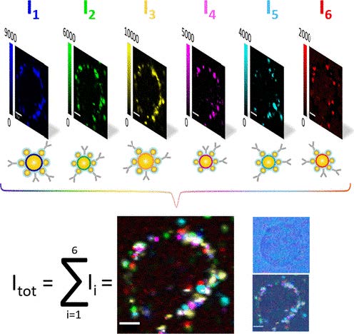
Surface-enhanced Raman scattering (SERS) as a labeling technique has several advantages over traditional staining methods such as immunohistochemistry [1]. One of them is the possibility to simultaneously localize multiple target proteins on cells and tissues for cancer diagnostics. Current bioimaging methods lack the necessary multicolor capacity and suffer from background/autofluorescence. Both these central limitations can be overcome by immuno-SERS microscopy using SERS nanotags conjugated to antibodies. We present a proof of concept study for the detection of a single target protein using six different Raman reporter-encoded core-satellite SERS nanotags on single breast cancer cells [2]. The target-protein is human epidermal growth factor receptor 2 (HER2) which is a prominent breast cancer marker and is expressed on the cell membrane of breast cancer cells. As shown in Figure 1 below, the six different false-color iSERS images clearly indicate the high abundance of HER2 on the cell membrane. These data demonstrate that iSERS is a promising alternative to traditional immunohistochemistry and immunofluorescence techniques in clinical applications for the simultaneous analysis of a panel of biomarkers at the level of single cells.

Bibliography:
[1] Schlücker, S., Angew. Chem. Int. Ed., 2014, 53, 4756-95.
[2] Stepula, E., Wang, X.-P., Srivastav, S., König, M., Levermann, J., Kasimir-Bauer, S., Schlücker, S., ACS Appl. Mater. Interfaces, 2020, 12, 29, 32321–27.


