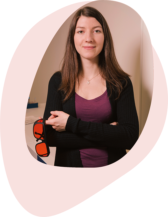
Judith S. Birkenfeld
Air-puff deformation imaging for early detection of ocular biomechanical changes
Institute of Optics “Daza de Valdés” // Madrid, Spain
Q&A Session III // Friday, October 30 // 8.15 am – 9 am (CET)

The cornea is the transparent, outer part of the eye. It is mainly composed of collagen, which gives it the microstructure that provides mechanical integrity necessary to maintain its typical dome-shape. Alterations in the cornea’s biomechanical properties, as they occur in corneal ectasia, lead to an abnormal corneal shape and a decreased quality of vision. Common types of corneal ectasia are keratoconus (KC) and surgically provoked ectasia. Diagnosis, management and treatment planning of KC currently depend on morphological measurements and biometric data, hence, corneal alterations are usually detected when refractive or topography changes have already occurred, and vision is irreversibly affected. Early detection of KC is key to introduce treatment options that can slow down the disease and lower the need for corneal transplants and subsequent re-grafts.
Corneal alterations are closely connected to the biomechanical properties of the cornea, and it has been recognized that the measurement of these properties help to diagnose tissue abnormalities. Currently, non-contact approaches that are investigated include OCT-Elastography, Brillouin microscopy, and Air-puff deformation imaging, whereby only the latter has been commercialized for the quantification of corneal biomechanics.
In our pitch, we will present recent approaches from the Visual Optics and Biophotonics Lab for the early detection of biomechanical changes in ocular tissue. We will introduce a custom‑developed swept-source optical coherence tomography (SSOCT) system, coupled with an air-puff excitation source that acquires dynamic deformation images on multiple meridians. We will show ex vivo and in vivo results of corneal and scleral deformation imaging, backed by advanced numerical simulations. We will further talk about effective data analysis tools that are based on the tissue’s deformation behavior in both, space and time. Finally, we will suggest helpful methods for the preliminary testing of new instruments that aim to quantify changes in ocular biomechanics, which include artificial and model corneas.


