Research Center for Applied Sciences, Academia Sinica, Nangang, Taipei, 115, Taiwan
Near Field Spectroscopic Imaging: from Hard to Soft Materials
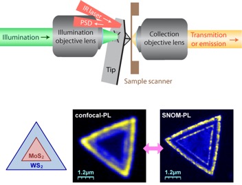
Scanning near field optical microscopy (SNOM) is a scanning probe technique that combines optics with an atomic force microscopy (AFM) to achieve sub-diffraction limit optical resolution. The tip-sample distance control is very critical and may causes serious sample damage during SNOM operation. This is still a mild issue for “hard” materials, such as transition metal dichalcogenides (TMD). Even though small scratch may happen, the sample would not be shifted by the tip. However, for “soft” materials, such as polymers and lipids, dragging and lifting of the sample are deadly problems to damage both the sample and SNOM tip.
Here we design and build a horizontal-type aperture based SNOM setup (a-SNOM) with superior mechanical stability toward high resolution and non-destructive topographic and optical imaging. We adopt the torsional resonance (TR) mode for the AFM operation to achieve a better force sensitivity and a higher spatial resolution even with the blunt a-SNOM tip. In addition, we developed another homemade horizontal-type SNOM system in an Argon purged glove box for stable SNOM operation and to eliminate possible photoreactions. Finally, a-SNOM imaging of fluorescent dye-labeled lipid domains are successfully achieved without sample damage by our horizontal-type a-SNOM system. In this talk, we will present simultaneous SNOM fluorescence and topographic imaging of TMD heterojunction, self-assembled P3HT nanowires and lipid domains with lateral optical resolution of 60~80 nm.
